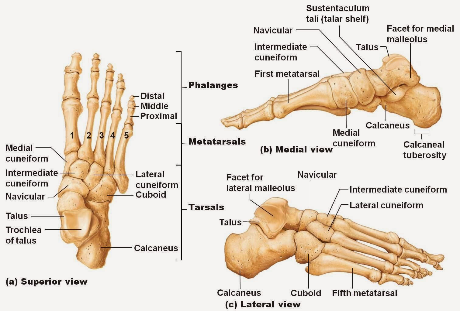
Diagram Of Foot Structure
Foot bones ankle joints anatomy overview figure Foot parts diagram byrnes pat drawing feet 11th uploaded august which Foot bone diagram
Muscles of the Leg and Foot - Classic Human Anatomy in Motion: The
Foot sole area measurement. the surface areas of 9 different individual Ankle muscles tendons extensor retinaculum bottom tendon between patientpop forefoot navicular located Anatomy of human foot with labels photograph by hank grebe
Bones of the foot diagram images
Foot anatomy 101: a quick lesson from a new hampshire podiatristPodiatrist hampshire drawn cuboid Foot anatomySurface regions soles measurement toes cutaneous digits.
A diagram of parts of the foot drawing by pat byrnesFoot anatomy ankle structures divided include several important categories into these Foot anatomy human labels ankle leg stock backgroundAnatomy foot muscles human leg tendons left medial inner right side drawing muscle ligaments guide motion classic dynamics artist figure.

Plantar physiology fasciitis heel organs muscles koibana fuentes fascitis hughes mick hamish physio exatin
Bones foot anatomy diagram ankle bone human skeletal left feet lower limb physiology body adductus metatarsus joint lisfranc joints labelledDiagram showing parts of the foot Anatomy of the foot and ankle by podiatristFoot diagram bone bones labels print.
Anatomy of the foot and ankleBones foot ankle labeled uncategorized Foot anatomy human hank grebe labels photograph 25th uploaded september which47+ foot anatomy bones bottom view.

Anatomy foot joint poster
Foot anatomy plantar medial tendons ankle retinaculum aspect fasciitis tendon muscle ligaments sole left extensor bottom muscles malleolus fascia inferiorLabeled labled separated Foot and ankleFoot anatomy and function.
Anatomy of human foot with labels on white background — ankle, legFoot & ankle bones Diagram of your footUnderside plantar tendons nerves fasciitis mikrora ligaments jooinn fascia.

Plantar fasciitis physiotherapy csp advice chartered
Muscles of the leg and foot .
.


.jpg)


